Childhood Cancer Info
Commonly Used Terms
Below are some of the terms commonly used when talking about the treatment of childhood cancer. Click on the term to reveal the explanation.
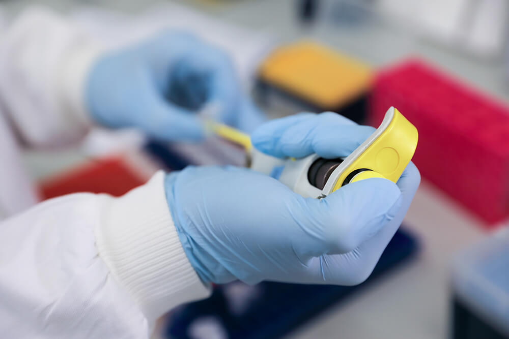
RESEARCH PROJECTS
Cancer is the leading cause of death in children aged 1-14 years in the UK and survivors can face a lifetime of serious health issues as a result of the intensive treatments used to treat their cancer. Childhood cancers are different to the cancers that occur in adults – dedicated research is needed. Would you like to help?
Related topics
We have lots of information to help you learn more about childhood cancer. From specific cancer types, to treatments and causes.
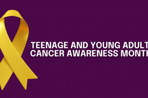
Teenage and Young Adult Cancer Awareness Month 2026
Throughout April, we’ll be sharing stories from young people about the impact of cancer on fertility, body image, mental health and education, to amplify their voices and raise awareness of their experiences.
Read more Teenage and Young Adult Cancer Awareness Month 2026
Mother’s Day
Mother’s Day We recently told you Leah’s story. After being told that her body would never be able to carry…
Read more Mother’s Day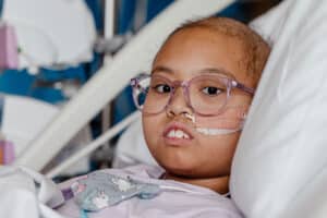
National Cancer Plan
National Cancer Plan: A welcome vision born from tragedy, but families need support today 4 February 2026 The National Cancer…
Read more National Cancer Plan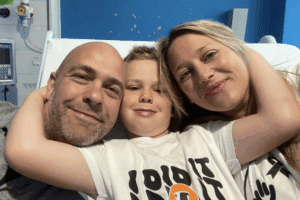
World Cancer Day 2026
World Cancer Day 2026 What is World Cancer Day? World Cancer Day takes place on February 4th every year. The…
Read more World Cancer Day 2026
More Child Research is Needed
2 Children received a terminal cancer diagnosis today. In 2026, this should’t happen. Only around 2% of cancer research funding…
Read more More Child Research is Needed
Childhood Cancer Research
Real research. Real breakthroughs. Real children who’ll benefit. Every contribution brings us one step closer to better young person’s treatments,…
Read more Childhood Cancer Research
Thank you for your support
Surviving childhood cancer is just the beginning Every contribution brings us one step closer to better young person’s treatments, improved…
Read more Thank you for your support
Childhood Cancer Treatment
Our children deserve cancer treatments designed for them. Every contribution brings us one step closer to more effective young person’s…
Read more Childhood Cancer Treatment
Childhood Cancer Report
FREE Childhood Cancer Report Only around 2% of cancer research funding in the UK* is spent on research that is…
Read more Childhood Cancer Report
Our Annual Reports
OUR ANNUAL REPORTS Each year we publish an impact report highlighting key areas of our work within childhood cancer research….
Read more Our Annual Reports